

id int64 56.3k 16.6M | text stringlengths 71 1.58k | t stringclasses 7
values | n stringclasses 4
values | m stringclasses 3
values |
|---|---|---|---|---|
56,344 | Complete atelectasis of the left upper lobe.
74 mm mass with poor contrast enhancement, obstructing the left upper lobe bronchus, considered to be lung cancer.
Enlargement of the left pulmonary hilum and ipsilateral mediastinal lymph nodes, suggesting lymph node metastases.
Enlarged lymph nodes on the right side of th... | T4 | N3 | M0 |
133,166 | Tumorous lesion with a diameter of 30 mm in the right lower lobe, S8. Its margin is spiculated. There is a possibility of lung cancer, so thorough examination in pulmonary medicine is recommended.
Cord-like opacity in the left lower lobe, similar to an inflammatory scar.
Enlargement of a right hilar lymph node is susp... | T1c | N0 | M0 |
165,742 | Pulmonary mass with a diameter of 5 cm in the right lung apex with spiculated margins, suspicious of lung cancer. Infiltration to the parietal pleura, suggesting T3.
No significant findings in the other lobes.
No mediastinal or hilar lymph node enlargement.
No pleural effusion.
The left adrenal gland is enlarged, like... | T3 | N0 | M0 |
404,886 | Tumorous lesion with a maximum diameter of 72 mm in the left lower lobe, suspicious of lung cancer.
The left lower lobe bronchus is infiltrated by the tumor, and there are infiltrative opacities around the tumor.
Associated obstructive pneumonia in the left lower lobe.
The left hilum and ipsilateral mediastinal lymp... | T4 | N2 | M1c |
463,397 | Prominent emphysematous changes. Irregular lobulated mass with a maximum diameter of 47 mm in the left upper lobe, with noticeable spiculated margins and pleural indentation. This correlates to a known lung cancer with pleural invasion. Hilar lymph nodes appear conglomerated. No other pathological lymph node enlargemen... | T2b | N0 | M0 |
493,069 | Solid mass with a diameter of 3 cm in the lower lobe of the left lung with spiculated margins, suspicious of primary lung cancer. A small nodule of 0.8 cm in size is seen within the same lobe, considered as T3.
No significant findings in the other lobes.
Enlargement of the left hilar lymph nodes, suspicious of metasta... | T3 | N1 | M1b |
862,683 | A 12 mm subpleural nodule in the right lower lobe, believed to be a finding of the known lung cancer. Interstitial pneumonia is suspected in both lungs. No pathological lymph node enlargement. No pleural effusion. Gallstones. | T1b | N0 | M0 |
906,857 | A solid mass of approximately 30 mm is observed in the lower lobe of the left lung, suspicious of lung cancer. There is a 9 mm nodule in its vicinity, and an 8 mm nodule in S8, suspicious of same-side intralobar metastases. Multiple soft tissue lesions spreading from the left pulmonary hilum to the mediastinum are susp... | T3 | N2 | M1c |
908,422 | An irregular mass of 35 mm is observed in the left lower lobe, suspicious of lung cancer. In addition, a nodular shadow is seen on the ventral side of the above lesion, possibly a pulmonary metastasis. Enlarged lymph nodes are observed in the left hilum, suggesting metastases. No pleural effusion. A hypovascular lesion... | T3 | N1 | M1b |
1,185,427 | A 30 mm diameter tumor is observed in the right lower lobe, suspicious of lung cancer. Accompanied by interlobar pleural indentation, visceral pleural invasion might be present. There are multiple focal ground-glass opacities in both upper lobes, recommended for follow-up. No significant lymph node enlargement in the h... | T1c | N0 | M0 |
1,538,432 | There is a tumor with a maximum diameter of 47 mm in the upper left lobe. It has infiltrated the lower lobe, crossing the interlobar pleura. The hilar lymph nodes are fused with the tumor. No enlargement of the mediastinal lymph nodes is observed. There is no pleural effusion. No liver or adrenal metastasis is observed... | T2b | N1 | M0 |
1,679,413 | Emphysematous changes.
A 47 mm irregular mass in the left upper lobe, suspicious of lung cancer. Possible invasion into the left pulmonary artery, as far as can be assessed with CT to a limited extent. The mass is also close to the aortic arch.
A small nodule is present in the right middle lobe, possibly inflammatory o... | T2b | N0 | M0 |
1,736,655 | S/P resection of right breast cancer. No sign of local recurrent tumor. Multiple masses are scattered in the mediastinum and right hilum. Lymph node metastasis is suspected. Differential diagnosis includes right hilar lung cancer and lymph node metastasis. Subpleural nodules in the right upper lobe, possibly metastases... | T3 | N2 | M0 |
1,856,176 | A tumor with a major axis of 1 cm, which obstructs the airways from the right main bronchus to the lower lobe bronchus, is observed at the right hilum, suspecting known lung cancer. Possible fusion of the mass with right hilar and subcarinal lymph nodes.
Atelectasis of the right lower lobe. There is a possibility that... | T2a | N2 | M0 |
1,863,157 | A nodule measuring 12 mm in diameter in the right lower lobe S8/9, increasing in size, suspected to be a known lung cancer. Reticular opacities present in the right S6, likely inflammatory changes. Calcified pleural plaques on both sides, suggesting the possibility of asbestos-related disease. No significant enlargeme... | T1b | N0 | M0 |
1,876,861 | Signs of chronic pulmonary emphysema.
An irregular tumor with a long diameter of 48 mm in the right lung apex, suspicious of primary lung cancer.
It is in contact with the pleura of the right lung apex, suspecting visceral pleural invasion.
No secondary tumor nodules.
Several enlarged hilar and mediastinal lymph node... | T2b | N2 | M1b |
1,956,050 | Emphysematous changes.
A mass is observed in the center of the left upper lobe. The left upper lobe bronchus is interrupted proximally, and irregularity and narrowing are observed in the left pulmonary artery and vein, which are partially interrupted. The mass corresponds to the known lung cancer.
The left upper lobe i... | T4 | N2 | M0 |
2,021,673 | A mass, approximately 12 cm in diameter, is observed primarily in the upper lobe of the left lung, extending across the interlobar pleura to the lower lobe of the left lung. The tumor is widely in contact with the mediastinum and pleura. Osteolytic changes are observed in the left third rib, suggesting infiltration. En... | T4 | N2 | M0 |
2,195,733 | Findings of chronic pulmonary emphysema. An irregular tumor with a long diameter of 35 mm in the left lower lobe S6, suspicious of primary lung cancer. Small nodules suspected of being peritumoral nodules are observed around the tumor. Enlargement of the left hilar lymph nodes, suspicious of metastasis. No pleural effu... | T3 | N1 | M1b |
2,240,304 | 46 mm mass in the right upper lobe, suspicious of lung cancer. The tumor protrudes into the subpleural adipose tissue, suggesting pleural invasion on the parietal side.
Enlargement of the mediastinal lymph nodes on the same side. Although not significant in size, the lymph nodes on the opposite side are also slightly ... | T3 | N2 | M1b |
2,254,357 | A tumor with a diameter of 12 cm is observed spreading across the upper and lower lobe of the left lung, corresponding to known lung cancer. It is in extensive contact with the pleura and is accompanied by the destruction of the left 3rd rib. Rib and parietal pleural infiltration is suspected. There are small nodules i... | T4 | N3 | M0 |
2,298,369 | A nodule protruding into the lumen of the right main bronchus and the intermediate bronchus, considered to be a known cancer lesion. The longest diameter is 17 mm. Due to this, the right middle lobe bronchus and right lower lobe bronchus are obstructed, and the right middle and lower lobes of the lung are collapsed. No... | T2a | N0 | M0 |
2,343,928 | No comparable imaging studies available. A lobulated tumor mass, approximately 47 mm in diameter, in the hilum of the left upper lobe, which is considered to be a known finding of lung cancer. The lesion appears to surround the bronchus of the left upper lobe, suggesting bronchial stenosis. Expansion into the mediastin... | T4 | N0 | M0 |
2,538,844 | A large tumorous lesion (primary focus 12 cm), centered in the upper lobe of the left lung. There is extensive contact with the visceral pleura, and pleural invasion is suspected. Some subpleural adipose tissue is irregular, and invasion into the chest wall is also suspected. The cortical bone of the left third rib is ... | T4 | N2 | M0 |
2,932,043 | A mass of approximately 74 mm is observed in the upper lobe of the left lung, not containing any air. Primary lung cancer is suspected. The central bronchus is obstructed by the tumor, leaving the upper lobe of the left lung in an almost completely atelectatic state.
No pulmonary nodules. There are no findings suggest... | T4 | N1 | M0 |
2,987,117 | Emphysema.
A mass with a maximum diameter of 4.7 cm in the upper lobe of the left lung. The tumor has a lobulated shape and spiculated margins, suggesting primary lung cancer. Infiltration into the mediastinal fat is observed, raising suspicion for T4.
No tumorous lesions in other lung fields.
On non-contrast CT, no pa... | T4 | N0 | M0 |
3,029,476 | A lobulated tumor with a diameter of 40 mm in the right lower lobe, suspicious of primary lung cancer. There is bronchiectasis with mucus retention below the tumor. Ground-glass opacities in the right upper lobe, likely inflammatory.
Enlargement of the right hilar and mediastinal lymph nodes, suspicious of metastasis.
... | T2a | N2 | M1c |
3,072,861 | A lobulated mass containg a cavity of 30 mm in diameter in the lower lobe of the left lung. Traction of the interlobar fissure and a spiculated margin are also observed. The mass is considered to be the known lung cancer. A secondary tumor nodule is suspected within the same lobe.
The left hilar lymph node is enlarged.... | T3 | N0 | M0 |
3,384,646 | A mass with a maximum diameter of 7.2 cm in the lower lobe of the left lung, suspicious of lung cancer. It is considered T4.
Stenosis of the airway caused by the tumor, and an increased density in the peripheral lung field, suspecting obstructive pneumonia.
Thickening of the pleura adjacent to the tumor and a small amo... | T4 | N2 | M1c |
3,599,413 | There is a 17 mm protruding lesion in the right bronchial lumen.
It is considered to be the known lesion.
The right upper lobe bronchus is patent, but the bronchus intermedius is obstructed by the tumor. Atelectasis is evident in the right middle and lower lobes.
There is a small amount of pleural effusion on the righ... | T2a | N2 | M0 |
4,092,235 | A 12 mm nodule is observed in the right lower lobe, which has increased in size compared to the CT in August 2012, possibly lung cancer.
Ground-glass opacities and reticular opacities are seen mainly in the subpleural area of the lower lung field, suggesting interstitial pneumonia.
No significant lymph node enlargemen... | T1b | N0 | M0 |
4,166,189 | A tumor with a major axis of 12 cm in the left upper lobe of the lung. Infiltration of the aortic arch and pulmonary artery is suspected. This is considered T4.
Enlargement of the left pulmonary hilum and a mediastinal lymph node in level 6 is observed, suspicious of metastases. This is considered N2.
No pleural effusi... | T4 | N2 | M0 |
4,465,068 | No comparable imaging studies are available.
There are multiple enlarged lymph nodes with heterogeneous enhancement in the right hilum and mediastinum. S/P resection of right breast cancer, but there is no significant lymph node enlargement in other areas, suggesting lymph node metastasis of small cell lung cancer.
Nod... | T0 | N2 | M0 |
4,624,494 | There is an irregular mass with a diameter of 50 mm in the apex of the right lung. It appears to protrude into the subpleural adipose tissue near the origin of the right second rib, and pleural invasion on the visceral side may be present. The right hilum and mediastinal lymph nodes are slightly enlarged, but not of si... | T2b | N0 | M0 |
4,644,367 | Mass of 72 mm in the left lower lobe, suspicious of lung cancer. Infiltratative opacities and ground-glass opacities are seen on the peripheral side, suggesting obstructive pneumonia. Enlarged lymph nodes in the mediastinum, above the left clavicle, and in the left axilla, suggesting metastases. Small amount of left pl... | T4 | N3 | M1c |
4,646,850 | Pulmonary emphysematous changes.
There is an irregularly margined mass in the lower lobe of the left lung, approximately 35 mm in diameter, considered to be the known lung cancer.
Apart from this tumor, a nodule of 8 mm in diameter is present in the left lower lobe, suspicious of metastasis.
No other lesions suggest... | T3 | N1 | M1c |
4,760,374 | A lobulated tumor mass with a diameter of 40 mm is present in segment S6 of the right lower lobe of the lung, suspicious of lung cancer. A ground-glass nodule with lobulated margins is present in segment S2 of the right upper lobe, which may also be lung cancer (adenocarcinoma in situ).
Emphysematous changes in the lun... | T2a | N2 | M1c |
4,844,803 | Moderate emphysematous changes in the lungs.
An irregularly margined tumor is observed in the central part of the left upper lung lobe. The margins are spiculated and the tumor has a maximum diameter of 47 mm. It is considered to be the known lung cancer. The tumor is in broad contact with the interlobar pleura.
The l... | T2b | N0 | M0 |
4,850,448 | Mild emphysematous changes of the lungs are present.
The left upper lobe is atelectatic. An irregular soft tissue mass is located centrally, suggesting the possibility of lung cancer. There is a possibility of infiltration into the mediastinum.
Left hilar lymph nodes are enlarged, suggesting the possibility of metasta... | T4 | N2 | M0 |
5,033,297 | No comparable imaging studies are available.
A lobulated tumor mass, approximately 30 mm in diameter is present in S6 of the left lower lobe of the lung. This is considered lung adenocarcinoma. A nodule of about 9 mm in diameter is in contact with the lesion, and a nodule of 8 mm in contact with the interlobar pleura ... | T3 | N1 | M1b |
5,073,667 | There is a solid tumor of 30 mm in S8 of the right lower lung lobe. It has a lobulated shape and is accompanied by pleural indentation. No sign of invasion beyond the interlobar pleura. No pulmonary metastasis. There is a small nodule in the apex of the right lung, follow up recommended. No pleural effusion. No gross a... | T1c | N0 | M0 |
5,582,789 | A soft tissue mass is present in the right hilum, suspected to be a conglomerate of lung cancer and hilar lymph node metastasis. Size evaluation is difficult. The mass is in contact with the tracheal bifurcation and major blood vessels, suggesting invasion. It is considered T4.
A small round subpleural nodule is presen... | T4 | N2 | M0 |
6,231,900 | A tumor-like lesion with irregular margins, measuring up to 72 mm in diameter, is present on the mediastinal side of the left lower lobe of the lung. The lesion shows heterogenous contrast enhancement. It is considered to correlate to the known cancer. The tumor is widely in contact with the pleura, but without clear m... | T4 | N2 | M1c |
6,232,953 | No comparable imaging studies are available.
A tumor of approximately 17 mm in diameter is observed at the right main bronchus. The bronchial lumen is narrowed and there is atelectasis peripheral thereof.
Soft tissue lesions, which are considered to be enlarged lymph nodes, are observed under the right hilum and the ... | T2a | N2 | M0 |
6,363,776 | Numerous tumorous lesions spreading from the right pulmonary hilum to the mediastinum, suspected to be small cell carcionma with lymph node enlargement. A nodule is present in the peripheral S3 of the right upper lobe, suspicious of pulmonary metastasis. In addition, there are scattered fine granular opacities, also su... | T3 | N2 | M0 |
6,441,303 | Emphysematous changes are prominent in the lungs.
There is a lobulated tumor of 40 mm in diameter in the right lower lobe, suggesting lung cancer. Multiple nodules and increased opacity along the airway in the surrounding area suggest obstructive pneumonia or metastatic nodules.
Ground-glass nodules are scattered in ... | T2a | N2 | M1c |
6,574,876 | There are multiple enlarged lymph nodes in the right mediastinum and right hilum, consistent with known small cell carcinoma. A small nodule is observed in the right upper lobe S3, possibly a metastatic nodule.
There is mild interstitial opacitiy in the peripheral lung, possibly interstitial pneumonia.
No pleural eff... | T3 | N2 | M0 |
6,778,649 | A tumor is present in the left pulmonary hilum, suggesting lung cancer. The right upper lobe bronchus is obstructed due to tumor infiltration, resulting in atelectasis of the left upper lobe. The border between the tumor and the atelectasis is difficult to evaluate. The left pulmonary artery is in contact with the tumo... | T4 | N3 | M0 |
6,793,001 | A 12 cm large tumor is present in the left lung apex, suggesting lung cancer. Destruction of the left 3rd rib suggests infiltration. No significant lymph node enlargement in the mediastinum. There is no pleural effusion. | T4 | N0 | M0 |
6,848,850 | Emphysema in both lungs.
A lobulated tumorous lesion is present in the lower lobe of the right lung (maximum diameter 37×37×40mm). Primary lung cancer is suspected. Soft tissue masses are found in the bronchi. Bronchial mucus plugs are observed.
Faint ground-glass opacities are scattered in both lungs.
Lymph node enlar... | T2a | N2 | M1c |
6,861,476 | There is a tumor with a major axis of 72 mm in the left lower lobe, suspicious of lung cancer. The tumor protrudes into the mediastinum and is in contact with the descending thoracic aorta, suggesting mediastinal invasion. There is interstitial thickening around the tumor, possibly carcinomatous lymphangitis.
There is ... | T4 | N2 | M1c |
7,127,615 | There is a tumor with a long diameter of 50 mm in the right lung apex with spiculated margins. The morphology suggests lung cancer. Emphysematous changes in both lungs.
No pathological lymph node enlargement. No pleural effusion.
No coarse lesions in the upper abdomen. | T2b | N0 | M0 |
7,245,712 | A tumor that has become a conglomerate with the hilar lymph nodes is seen on the central side of the left upper lobe. The left upper lobe bronchus is obstructed, and atelectasis is present, making it difficult to measure the size of the tumor. The tumor protrudes into the mediastinum, suggesting mediastinal invasion.
T... | T4 | N2 | M0 |
7,467,648 | An irregular mass of 30 mm in size is observed in the right lower lobe, considered to be the suspected lung cancer.
A pure GGN is present in the left lung apex, which should be checked in follow-up.
Striated opacities suspicious of inflammatory scars are seen in both lower lobes.
No significant lymph node enlargemen... | T1c | N0 | M0 |
7,516,987 | There is a small subpleural nodule in the right upper lobe of the lung, S3, possibly lung cancer.
There are subpleural reticular opacities of both lungs, suspecting interstitial pneumonia.
The right hilar, ipsilateral, and contralateral mediastinal lymph nodes are enlarged, considered to be lymph node metastasis.
No pl... | T0 | N3 | M0 |
7,547,062 | Compared with the CT from August 2012.
A nodule of about 12 mm in diameter is located in S9 of the right lower lobe. In the CT from August 2012, a focal ground-glass opacity was seen in the same area, which seems to be progressing. Lung cancer is known, and this lesion is considered cancerous based on the progression.
... | T1b | N0 | M0 |
7,616,690 | Emphysema is observed.
Subpleural reticular opacities and ground-glass opacities are seen in both lower lobes, suggesting interstitial changes.
There is a solid subpleural tumor with a long diameter of 1.2 cm of the right lower lobe. Compared to the previous examination, it has increased in size, suggesting primary lun... | T1b | N0 | M0 |
7,656,084 | No comparable imaging studies are available.
A tumor mass measuring 12 cm in diameter is observed in the left lung apex. The tumor extends to the chest wall and ribs, accompanied by bone destruction. This is considered indicative of lung cancer with chest wall invasion.
No obvious metastasis or other active lesions i... | T4 | N2 | M0 |
7,751,255 | No comparable imaging studies are available. A lobulated tumor mass, approximately 30 mm in diameter, is present in S8 of the right lower lobe of the lung, accompanied by a slight ground-glass opacity in its periphery. This is considered to be a finding of known lung cancer. Multiple pure GGNs up to about 10 mm are fou... | T1c | N0 | M0 |
7,793,337 | A nodular infiltratative opacity of 30 mm in diameter is present in the right lower lobe S8, corresponding to known lung cancer. It is in extensive contact with the interlobar pleura, suggesting infiltration. No findings suspicious of metastasic nodules are observed in both lungs. Striated opacities are present in both... | T2a | N0 | M0 |
7,943,636 | A nodular mass measuring 1.7 cm in diameter is found in the right main bronchus. No lesions are seen in the bronchial bifurcation, thus the tumor is considered T2. Obstructive atelectasis is present. No significant findings in other lung segments. Lymph nodes in the left supraclavicular fossa are enlarged, suspecting N... | T2a | N3 | M0 |
8,100,850 | There is a nodule measuring 17 mm in diameter in the right main bronchus, which correlates to the known mucinous squamous cell carcinoma. The right lower lobe bronchus is obstructed. The right lower lobe is collapsed. A small amount of pleural effusion on the right. No secondary tumor nodules in the ventilated lung. Th... | T2a | N3 | M0 |
8,221,825 | A tumor with a major axis of 12 cm is located in the upper and lower lobe of the left lung, suspecting lung cancer. The tumor is destroying the third rib and infiltrating the chest wall. Also, mediastinal infiltration is suspected, and it is in close proximity to the aortic arch. There is a small nodule in the peripher... | T4 | N2 | M0 |
8,235,226 | Moderate chronic pulmonary emphysema. An irregular tumor with a long diameter of 47 mm is seen in the left upper lobe at the pulmonary hilum, suspicious primary lung cancer. It is in extensive contact with the interlobar pleura, suggesting infiltration. Mediastinal fat infiltration is also suspected. No significant enl... | T4 | N0 | M0 |
8,602,016 | A soft tissue mass in the right hilum correlating to the suspected malignant tumor. The right lower lobe is atelectatic.
A nodule is seen above the left clavicle, possibly lymph node metastasis.
A small amount of pleural effusion on the right. | T2a | N3 | M0 |
8,760,223 | There is a lobulated tumor with a maximum diameter of 40 mm in S6 of the right lower lobe. It is surrounded by a ground-glass opacities, and it is considered to be lung cancer. There is a tubular structure continuous with the tumor, and its lumen is enhanced. There is a possibility that the tumor has infiltrated the br... | T2a | N0 | M1c |
8,811,397 | There is a lobulated irregular tumor measuring 46 mm in the right lung apex, which is considered to be known of lung cancer. The right mediastinal lymph nodes are enlarged, suggesting metastasis. The left adrenal gland is significantly enlarged, possibly metastasis. No pleural effusion. | T2b | N2 | M1b |
9,175,672 | Enlarged lymph nodes in the right pulmonary hilum and the mediastinum, raising suspicion for lung cancer. Small lymph nodes can also be seen in the left mediastinum, but their size is not significant.
There is a nodule in the right upper lobe, considered to be a secondary tumor nodule within the same lobe.
Ground-glas... | T3 | N2 | M0 |
9,262,204 | There is a 12 mm subpleural nodule of the right lower lobe in S9, surrounded by ground-glass opacity. Compared to the CT in August 2012, it has increased in size, and we suspect lung cancer. There are subpleural reticular opacities and ground-glass opacities in both lungs, consistent with interstitial pneumonia. There ... | T1b | N0 | M0 |
9,524,486 | Emphysema and interstitial changes in both lungs. Thickening and calcification of the pleura on both sides. Asbestosis seems to be present.
An irregular subpleural nodule is found in the right lung in S9 (approximately 12 mm), possibly primary lung cancer associated with asbestosis.
Multiple mediastinal lymph nodes, bu... | T1b | N0 | M0 |
9,635,688 | A dense ground-glass opacity measuring 30 mm in diameter is located in S8 of the right lower lung lobe. It is somewhat indistinct and partially lobulated. It is considered to be consistent with a cancerous lesion. No clear pleural invasion.
Multiple ground-glass opacities of several millimeters in both upper lung lobes... | T1c | N0 | M0 |
9,651,856 | Plaques with some coarse calcification are scattered on both pleurae, suggesting asbestos-related pleural disease. Mild emphysematous changes of the lung parenchyma. Ground-glass opacities and reticular opacities are seen in the peripheral lung parenchyma, predominantly in the lower lobes on both sides, suggesting inte... | T1b | N0 | M0 |
9,673,524 | There is a lobulated tumor with a maximum diameter of 46 mm in the right lung apex. It is accompanied by a ground-glass opacity around it. The tumor is considered to be lung cancer. No pleural effusion. The right hilar, ipsilateral, and contralateral mediastinal lymph nodes are enlarged, suspicious of lymph node metast... | T2b | N3 | M1b |
9,745,005 | Emphysema.
There is a solid mass with a maximum diameter of 47 mm in the upper lobe of the left lung, without any air inside, suspicious of primary lung cancer. Invasion of the visceral pleura is suspected. There are no findings suggesting lung metastasis.
No pathological lymph node enlargement in the mediastinum or hi... | T2b | N0 | M0 |
9,818,606 | Emphysematous changes of the lung parenchyma.
A tumor with a diameter of 40 mm is located in the lower lobe of the right lung. It has a lobulated shape, and lung cancer is strongly suspected. There are multiple granular opacities and mucus plugs in the periphery, which are considered to be signs of obstructive pneumoni... | T2a | N2 | M1c |
9,905,822 | A tumor with a major axis of 4.6 cm is located in the right lung apex, surrounded by spiculated opacities. Primary lung cancer is suspected. Pleural invasion is present at the lung apex, considered as T3.
Bullae are scattered in the left upper lobe of the lung.
Enlargement of the mediastinal lymph nodes in level 4R, su... | T3 | N2 | M1b |
10,223,615 | A lobulated irregular mass of about 3 cm is located in the subpleural area of the peripheral right lung lower lobe in S8. The main component is solid, but ground-glass opacities can also be seen in its periphery. Pleural retraction is also observed. Primary lung cancer is suspected. | T1c | N0 | M0 |
10,320,785 | Multiple tumors are present, spreading from the right hilum to the mediastinum. It is considered to be known small cell carcinoma and its metastases. The size of the primary tumor is unknown.
There is a pleura-based nodule in the right upper lobe, suspicious of metastasis.
Reticular opacities are located in the periphe... | T3 | N2 | M0 |
10,405,379 | There is a nodule of 17 mm in diameter in the right main bronchus, which appears to be part of the known mucinous squamous cell carcinoma. The right lower lobe branch is obstructed, causing atelectasis of the lower lobe. Pleural effusion on the right. No pathological lymph node enlargement. No significant lesions in th... | T2a | N0 | M0 |
11,013,624 | There is a large 12 cm hypodense tumor in the left upper lobe, considered to be lung cancer. It is widely in contact with the pleura on the mediastinal side, and pleural invasion is suspected. There is no direct invasion to the mediastinum. The left pulmonary hilum and ipsilateral mediastinal lymph nodes are enlarged, ... | T4 | N2 | M0 |
11,044,030 | There is a noticeable chain-like lymph node enlargement in the mediastinum, which is believed to be a known lymph node lesion. A small nodule is observed in the right upper lobe. Could this be a primary lung cancer lesion? No pleural effusion. | T0 | N2 | M0 |
11,079,957 | A solid tumor-like lesion of approximately 72 mm is located in the center of the left lung's lower lobe, S10. Primary lung cancer is suspected. It is in contact with the pleura, and pleural invasion is suspected. Pleural effusion and pleural thickening are noted, suspecting pleural dissemination.
There are nodules in t... | T4 | N0 | M1c |
11,117,507 | There is a 50 mm neoplastic lesion with spiculated margins in the right lung apex. It is widely in contact with the pleura, and pleural invasion is suspected. There is no invasion to the ribs. Both lungs have subpleural reticular opacities and ground-glass opacities, suggesting interstitial pneumonia. No lung metastasi... | T2b | N0 | M0 |
11,328,933 | In the coronary reconstruction images, mixed ground-glass opacity (GGO) is observed in the right lower lobe of the lung, considered to be the known lung cancer. The solid part is 3 cm in diameter, which is considered T1c.
A ground-glass nodule is also seen in the apex of the left lung. Although small, it is nearly full... | T1c | N0 | M0 |
11,595,888 | A 50 mm tumor is located in the apex of the right lung, in contact with the pleura. The tumor has irregular margins and spiculated appearance. Osteolytic lesions are observed in the right second rib in contact with the tumor, suggesting infiltration. These findings strongly suggest lung cancer. Emphysematous changes ar... | T3 | N0 | M0 |
11,604,112 | Emphysematous changes of the lung parenchyma.
An irregular mass is located in the apex of the right lung, which is considered to be a known cancer lesion. Its longest diameter is 46 mm. It is widely in contact with the pleura, suggesting invasion. However, clear invasion into the chest wall cannot be pointed out.
No l... | T2b | N2 | M1b |
11,650,032 | There is an irregular tumor measuring 35 mm in the distal part of the left lower lobe, accompanied by retraction of the interlobar pleura. There is a nodule measuring 8 mm in the left lower lobe, suspected to be a metastatic nodule. Lymph nodes in the left hilum are enlarged, suggesting metastasis. No significant media... | T3 | N1 | M1c |
11,789,977 | No comparable imaging studies are available.
Atelectasis is observed in the left lingular segment and part of the upper lobe. No tumor can be seen centrally, but there is narrowing and obstruction of the airways from the left main bronchus to the peripheral bronchi, which is considered to be lung cancer accompanied by ... | T2a | N2 | M0 |
12,912,312 | No comparable imaging examinations are available.
A tumor mass of 72 mm in diameter is located in the lower lobe of the left lung. It is considered to correspond to known lung cancer. There are no obvious metastases in the lungs.
Emphysematous changes.
Pleural effusion on the left. The pleura is slightly thickened, sug... | T4 | N2 | M1c |
12,958,437 | Emphysematous changes and bullae in the lung parenchyma.
A tumorous lesion measuring 27 × 34 × 46 mm is observed in the right upper lobe of the lung. No intralesional air. Primary lung cancer is suspected. It is in contact with the pleura, and visceral pleural invasion is suspected.
The mediastinal lymph nodes are enl... | T2b | N2 | M1b |
13,029,131 | A tumor is located in the left pulmonary hilum. It protrudes into parts of the upper lobe bronchus, and the periphery has become alectatic. Towards the mediastinum, it is in contact with the heart and there is a possibility of invasion. It is considered T4.
Small nodules are scattered in the lower lobe of the right lun... | T4 | N1 | M0 |
13,358,309 | There is a tumor with a maximum diameter of 30 mm in the left lower lobe S6, and a focal ground-glass opacity of 9 mm in contact with the tumor.
In addition, there is a nodule of 8 mm along the interlobar pleura of the left lower lobe S6.
Lung cancer and pulmonary metastases are suspected.
The left hilar lymph nodes... | T3 | N1 | M1b |
13,567,317 | Interstitial pneumonia and pulmonary emphysema. A condition that is prone to the development of lung cancer, such as emphysema combined with pulmonary fibrosis, is suspected.
A tumorous lesion of approximately 50 mm is located mainly in the right upper lobe of the lung. Primary lung cancer is suspected.
Infiltration in... | T2b | N2 | M0 |
13,658,329 | There is suspicion of chronic pulmonary emphysema in the lung parenchyma. A tumor with a long diameter of 50 mm with spiculated margins and extensive contact to the pleura, is located in the right lung apex. Lung cancer with visceral pleural invasion is suspected. No obvious secondary tumor nodules in the lungs. No sig... | T2b | N0 | M0 |
14,133,008 | There is a 72 mm tumor in the left lower lobe, which is considered to be the primary focus of known lung cancer. Pleural effusion on the left. Pleural dissemination cannot be ruled out. The lymph nodes below the tracheal bifurcation are enlarged, suspicious of metastases. | T4 | N2 | M0 |
14,133,817 | Chest:
Emphysematous changes.
A 40 mm tumor is located in the right lower lobe, accompanied by pleural invasion. Lung cancer is suspected. A bronchial mucus plug is suspected below the tumor, but a tumor thrombus cannot be ruled out.
There is a faint ground-glass opacity in the right upper lobe, which may be an inflamm... | T2a | N1 | M1c |
15,005,039 | No comparable imaging studies are available.
A lobulated tumor mass, measuring 46 mm in the longest dimension, is observed in the right lung apex, which is considered to be a finding of known lung cancer.
Centrilobular emphysema is seen mainly in the upper lung. There are no obvious metastases or other active lesions... | T2b | N2 | M1b |
15,045,923 | A tumor shadow of 50 mm is located in the apex of the right lung with irregular shape and spiculated margins, suggesting lung cancer. It is widely in contact with the chest wall, suspecting infiltration. There are interstitial changes such as mild emphysematous changes mainly subpleural, reticular opacities, and ground... | T2b | N0 | M0 |
15,059,875 | There is a lobulated tumor with a long diameter of 4.0 cm in the right lower lobe of the lung, suspecting lung cancer. There are nodular opacities around the lesion, suspected to be obstructive atelectasis due to the tumor. It is considered T2.
Lymph node enlargement is seen on the right side of the trachea, suspecting... | T2a | N2 | M1c |
15,095,613 | Severe emphysematous changes. There is an irregular tumor of 47 mm in the upper lobe of the left lung, suspecting lung cancer. The tumor has narrowed the upper lobe bronchus. Also, the tumor is closely associated with the main trunk of the left pulmonary artery, raising suspicion of invasion. Please re-evaluate in cont... | T4 | N1 | M0 |
RadNLP 2024 main task: Document Classification for Lung Cancer Staging

📚 Introduction
RadNLP 2024 is a shared task in the international conference NTCIR-18, organized by the National Institute of Informatics in Japan.
Management of lung cancer is based on the stage, and radiology reports provide various related information by describing medical images such as CT and MRI.
However, radiology reports do not always specify the stage explicitly¹. This imposes extra workload on human experts for careful manual information extraction, which can be aided by automation.
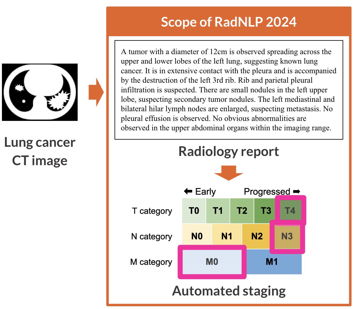
Main task is a multi-label document classification to correctly determine T, N, and M categories for each radiology report.
- T category - Assessment of the size and/or extension of the primary lesion. Available choices: T0, Tis, T1mi, T1a, T1b, T1c, T2a, T2b, T3, T4
- N category - Assessment of the extent of lymph node metastasis. Available choices: N0, N1, N2, N3
- M category - Assessment of the extent of distant metastasis. Available choices: M0, M1a, M1b, M1c
In RadNLP 2024, we follow the staging criteria by the Japan Lung Cancer Society (JLCS). Note that the JLCS criteria closely aligns the global standard, namely the 8th edition of the TNM Classification of Malignant Tumours by the Union for International Cancer Control (UICC).
All radiology reports in RadNLP 2024 diagnose lung cancer at the initial evaluation. There are no reports of lung cancer post-treatment evaluation or different cancer.
Our datasets contain NO personal health information. The radiology reports are not derived from real medical institutions but are created with crowdsourcing by diagnosing de-identified images on Radiopaedia. Task participants requires no complex applications to use our datasets.
📊 Dataset Size & Format
This repository contains 6 files:
| Language | Split | Number of radiology reports |
|---|---|---|
| English | Training | 108 |
| English | Validation | 54 |
| English | Test | 81 |
| English | Total | 243 |
| Japanese | Training | 108 |
| Japanese | Validation | 54 |
| Japanese | Test | 216 |
| Japanese | Total | 378 |
Each file contains 5 columns:
| Column | Type | Description |
|---|---|---|
id |
int | Unique identifier |
text |
string | Radiology Report |
t |
string | Gold standard for the T category |
n |
string | Gold standard for the N category |
m |
string | Gold standard for the M category |
📖 Citation
If you find our work useful, please consider citing:
@inproceedings{radnlp2024,
  title={NTCIR-18 RadNLP 2024 Overview: Dataset and Solutions for Automated Lung Cancer Staging},
  author={Nakamura, Yuta and Fujimoto, Koji and Kluckert, Jonas and Krauthammer, Michael and Kanzawa, Jun and Katayama, Akira and Kikuchi, Tomohiro and Kurokawa, Ryo and Gonoi, Wataru and Tashiro, Yuki and Hanaoka, Shouhei and Yada, Shuntaro and Aramaki, Eiji},
  year={2025},
  booktitle={NTCIR-18: Proceedings of the 18th NTCIR Conference on Evaluation ofInformation Access Technologies},
  doi={10.20736/0002002061}
}
📜 License
This project is licensed under the CC-BY-4.0 license.
- Downloads last month
- 33