
Datasets:

id int64 | sentence_index int64 | text string | omittable int64 | measure int64 | extension int64 | atelectasis int64 | satellite int64 | lymphadenopathy int64 | pleural int64 | distant int64 |
|---|---|---|---|---|---|---|---|---|---|---|
56,344 | 0 | Complete atelectasis of the left upper lobe.
| 0 | 0 | 0 | 1 | 0 | 0 | 0 | 0 |
56,344 | 1 | 74 mm mass with poor contrast enhancement, obstructing the left upper lobe bronchus, considered to be lung cancer.
| 0 | 1 | 1 | 0 | 0 | 0 | 0 | 0 |
56,344 | 2 | Enlargement of the left pulmonary hilum and ipsilateral mediastinal lymph nodes, suggesting lymph node metastases.
| 0 | 0 | 0 | 0 | 0 | 1 | 0 | 0 |
56,344 | 3 | Enlarged lymph nodes on the right side of the trachea, also suspected to be lymph node metastases. | 0 | 0 | 0 | 0 | 0 | 1 | 0 | 0 |
56,344 | 4 | The left lower lobe bronchus is also infiltrated by the tumor and is narrowed.
| 0 | 0 | 1 | 0 | 0 | 0 | 0 | 0 |
56,344 | 5 | No pleural effusion.
| 1 | 0 | 0 | 0 | 0 | 0 | 0 | 0 |
56,344 | 6 | Normal appearance of the visualized upper abdomen. | 1 | 0 | 0 | 0 | 0 | 0 | 0 | 0 |
133,166 | 0 | Tumorous lesion with a diameter of 30 mm in the right lower lobe, S8. | 0 | 1 | 0 | 0 | 0 | 0 | 0 | 0 |
133,166 | 1 | Its margin is spiculated. | 1 | 0 | 0 | 0 | 0 | 0 | 0 | 0 |
133,166 | 2 | There is a possibility of lung cancer, so thorough examination in pulmonary medicine is recommended.
| 0 | 1 | 0 | 0 | 0 | 0 | 0 | 0 |
133,166 | 3 | Cord-like opacity in the left lower lobe, similar to an inflammatory scar.
| 1 | 0 | 0 | 0 | 0 | 0 | 0 | 0 |
133,166 | 4 | Enlargement of a right hilar lymph node is suspected. | 0 | 0 | 0 | 0 | 0 | 1 | 0 | 0 |
133,166 | 5 | No pleural effusion. | 1 | 0 | 0 | 0 | 0 | 0 | 0 | 0 |
165,742 | 0 | Pulmonary mass with a diameter of 5 cm in the right lung apex with spiculated margins, suspicious of lung cancer. | 0 | 1 | 0 | 0 | 0 | 0 | 0 | 0 |
165,742 | 1 | Infiltration to the parietal pleura, suggesting T3.
| 0 | 1 | 1 | 0 | 0 | 0 | 0 | 0 |
165,742 | 2 | No significant findings in the other lobes.
| 1 | 0 | 0 | 0 | 0 | 0 | 0 | 0 |
165,742 | 3 | No mediastinal or hilar lymph node enlargement.
| 1 | 0 | 0 | 0 | 0 | 0 | 0 | 0 |
165,742 | 4 | No pleural effusion.
| 1 | 0 | 0 | 0 | 0 | 0 | 0 | 0 |
165,742 | 5 | The left adrenal gland is enlarged, likely an adenoma.
| 1 | 0 | 0 | 0 | 0 | 0 | 0 | 0 |
165,742 | 6 | No significant findings in other abdominal organs. | 1 | 0 | 0 | 0 | 0 | 0 | 0 | 0 |
404,886 | 0 | Tumorous lesion with a maximum diameter of 72 mm in the left lower lobe, suspicious of lung cancer.
| 0 | 1 | 0 | 0 | 0 | 0 | 0 | 0 |
404,886 | 1 | The left lower lobe bronchus is infiltrated by the tumor, and there are infiltrative opacities around the tumor.
| 0 | 0 | 1 | 1 | 0 | 0 | 0 | 0 |
404,886 | 2 | Associated obstructive pneumonia in the left lower lobe.
| 0 | 0 | 0 | 1 | 0 | 0 | 0 | 0 |
404,886 | 3 | The left hilum and ipsilateral mediastinal lymph nodes are enlarged, suspicious of lymph node metastases.
| 0 | 0 | 0 | 0 | 0 | 1 | 0 | 0 |
404,886 | 4 | The left axillary lymph nodes are enlarged, suspicious of lymph node metastases.
| 0 | 0 | 0 | 0 | 0 | 0 | 0 | 1 |
404,886 | 5 | Small amount of pleural effusion on the left.
| 0 | 0 | 0 | 0 | 0 | 0 | 1 | 0 |
404,886 | 6 | Osteolytic lesions in the proximal end of the left humerus and the left scapula, suspicious of bone metastases.
| 0 | 0 | 0 | 0 | 0 | 0 | 0 | 1 |
404,886 | 7 | Cystic lesion in the midline skin of the back, possibly a sebaceous cyst. | 1 | 0 | 0 | 0 | 0 | 0 | 0 | 0 |
463,397 | 0 | Prominent emphysematous changes. | 1 | 0 | 0 | 0 | 0 | 0 | 0 | 0 |
463,397 | 1 | Irregular lobulated mass with a maximum diameter of 47 mm in the left upper lobe, with noticeable spiculated margins and pleural indentation. | 0 | 1 | 1 | 0 | 0 | 0 | 0 | 0 |
463,397 | 2 | This correlates to a known lung cancer with pleural invasion. | 0 | 1 | 1 | 0 | 0 | 0 | 0 | 0 |
463,397 | 3 | Hilar lymph nodes appear conglomerated. | 0 | 0 | 0 | 0 | 0 | 1 | 0 | 0 |
463,397 | 4 | No other pathological lymph node enlargement. | 1 | 0 | 0 | 0 | 0 | 0 | 0 | 0 |
463,397 | 5 | No pleural effusion. | 1 | 0 | 0 | 0 | 0 | 0 | 0 | 0 |
493,069 | 0 | Solid mass with a diameter of 3 cm in the lower lobe of the left lung with spiculated margins, suspicious of primary lung cancer. | 0 | 1 | 0 | 0 | 0 | 0 | 0 | 0 |
493,069 | 1 | A small nodule of 0.8 cm in size is seen within the same lobe, considered as T3.
| 0 | 1 | 0 | 0 | 1 | 0 | 0 | 0 |
493,069 | 2 | No significant findings in the other lobes.
| 1 | 0 | 0 | 0 | 0 | 0 | 0 | 0 |
493,069 | 3 | Enlargement of the left hilar lymph nodes, suspicious of metastasis. | 0 | 0 | 0 | 0 | 0 | 1 | 0 | 0 |
493,069 | 4 | No lymph node metastasis in the mediastinum or on the opposite side, considered as N1.
| 0 | 0 | 0 | 0 | 0 | 1 | 0 | 0 |
493,069 | 5 | No pleural effusion.
| 1 | 0 | 0 | 0 | 0 | 0 | 0 | 0 |
493,069 | 6 | A small nodule in liver S5. | 0 | 0 | 0 | 0 | 0 | 0 | 0 | 1 |
493,069 | 7 | It is a single lesion, but there is a possibility of metastasis.
| 0 | 0 | 0 | 0 | 0 | 0 | 0 | 1 |
493,069 | 8 | Bilateral renal cysts.
| 1 | 0 | 0 | 0 | 0 | 0 | 0 | 0 |
493,069 | 9 | No significant findings in the gallbladder, pancreas, or spleen.
| 1 | 0 | 0 | 0 | 0 | 0 | 0 | 0 |
493,069 | 10 | No enlargement of the abdominal lymph nodes.
| 1 | 0 | 0 | 0 | 0 | 0 | 0 | 0 |
493,069 | 11 | No ascites. | 1 | 0 | 0 | 0 | 0 | 0 | 0 | 0 |
862,683 | 0 | A 12 mm subpleural nodule in the right lower lobe, believed to be a finding of the known lung cancer. | 0 | 1 | 0 | 0 | 0 | 0 | 0 | 0 |
862,683 | 1 | Interstitial pneumonia is suspected in both lungs. | 1 | 0 | 0 | 0 | 0 | 0 | 0 | 0 |
862,683 | 2 | No pathological lymph node enlargement. | 1 | 0 | 0 | 0 | 0 | 0 | 0 | 0 |
862,683 | 3 | No pleural effusion. | 1 | 0 | 0 | 0 | 0 | 0 | 0 | 0 |
862,683 | 4 | Gallstones. | 1 | 0 | 0 | 0 | 0 | 0 | 0 | 0 |
906,857 | 0 | A solid mass of approximately 30 mm is observed in the lower lobe of the left lung, suspicious of lung cancer. | 0 | 1 | 0 | 0 | 0 | 0 | 0 | 0 |
906,857 | 1 | There is a 9 mm nodule in its vicinity, and an 8 mm nodule in S8, suspicious of same-side intralobar metastases. | 0 | 0 | 0 | 0 | 1 | 0 | 0 | 0 |
906,857 | 2 | Multiple soft tissue lesions spreading from the left pulmonary hilum to the mediastinum are suspicious of lymph node metastases. | 0 | 0 | 0 | 0 | 0 | 1 | 0 | 0 |
906,857 | 3 | Multiple nodular lesions with ill-defined borders in the liver, suspicious of liver metastases. | 0 | 0 | 0 | 0 | 0 | 0 | 0 | 1 |
908,422 | 0 | An irregular mass of 35 mm is observed in the left lower lobe, suspicious of lung cancer. | 0 | 1 | 0 | 0 | 0 | 0 | 0 | 0 |
908,422 | 1 | In addition, a nodular shadow is seen on the ventral side of the above lesion, possibly a pulmonary metastasis. | 0 | 0 | 0 | 0 | 1 | 0 | 0 | 0 |
908,422 | 2 | Enlarged lymph nodes are observed in the left hilum, suggesting metastases. | 0 | 0 | 0 | 0 | 0 | 1 | 0 | 0 |
908,422 | 3 | No pleural effusion. | 1 | 0 | 0 | 0 | 0 | 0 | 0 | 0 |
908,422 | 4 | A hypovascular lesion is observed in the liver S5, which appears to be continuous with the gallbladder, suspicious of metastasis, or possibly gallbladder cancer. | 0 | 0 | 0 | 0 | 0 | 0 | 0 | 1 |
908,422 | 5 | Please evaluate in conjunction with clinical findings. | 0 | 0 | 0 | 0 | 0 | 0 | 0 | 1 |
908,422 | 6 | A cyst in the left kidney. | 1 | 0 | 0 | 0 | 0 | 0 | 0 | 0 |
908,422 | 7 | No ascites. | 1 | 0 | 0 | 0 | 0 | 0 | 0 | 0 |
1,185,427 | 0 | A 30 mm diameter tumor is observed in the right lower lobe, suspicious of lung cancer. | 0 | 1 | 0 | 0 | 0 | 0 | 0 | 0 |
1,185,427 | 1 | Accompanied by interlobar pleural indentation, visceral pleural invasion might be present. | 0 | 0 | 1 | 0 | 0 | 0 | 0 | 0 |
1,185,427 | 2 | There are multiple focal ground-glass opacities in both upper lobes, recommended for follow-up. | 0 | 0 | 0 | 0 | 1 | 0 | 0 | 0 |
1,185,427 | 3 | No significant lymph node enlargement in the hilum or mediastinum. | 1 | 0 | 0 | 0 | 0 | 0 | 0 | 0 |
1,185,427 | 4 | No pleural effusion. | 1 | 0 | 0 | 0 | 0 | 0 | 0 | 0 |
1,538,432 | 0 | There is a tumor with a maximum diameter of 47 mm in the upper left lobe. | 0 | 1 | 0 | 0 | 0 | 0 | 0 | 0 |
1,538,432 | 1 | It has infiltrated the lower lobe, crossing the interlobar pleura. | 0 | 0 | 1 | 0 | 0 | 0 | 0 | 0 |
1,538,432 | 2 | The hilar lymph nodes are fused with the tumor. | 0 | 0 | 0 | 0 | 0 | 1 | 0 | 0 |
1,538,432 | 3 | No enlargement of the mediastinal lymph nodes is observed. | 1 | 0 | 0 | 0 | 0 | 0 | 0 | 0 |
1,538,432 | 4 | There is no pleural effusion. | 1 | 0 | 0 | 0 | 0 | 0 | 0 | 0 |
1,538,432 | 5 | No liver or adrenal metastasis is observed. | 1 | 0 | 0 | 0 | 0 | 0 | 0 | 0 |
1,538,432 | 6 | No significant abnormalities are observed in the visualized abdominal organs. | 1 | 0 | 0 | 0 | 0 | 0 | 0 | 0 |
1,679,413 | 0 | Emphysematous changes.
| 1 | 0 | 0 | 0 | 0 | 0 | 0 | 0 |
1,679,413 | 1 | A 47 mm irregular mass in the left upper lobe, suspicious of lung cancer. | 0 | 1 | 0 | 0 | 0 | 0 | 0 | 0 |
1,679,413 | 2 | Possible invasion into the left pulmonary artery, as far as can be assessed with CT to a limited extent. | 0 | 0 | 1 | 0 | 0 | 0 | 0 | 0 |
1,679,413 | 3 | The mass is also close to the aortic arch.
| 0 | 0 | 1 | 0 | 0 | 0 | 0 | 0 |
1,679,413 | 4 | A small nodule is present in the right middle lobe, possibly inflammatory or metastasis, re-evaluation recommended in follow-up.
| 0 | 0 | 0 | 0 | 1 | 0 | 0 | 0 |
1,679,413 | 5 | No significant mediastinal lymph node enlargement.
| 1 | 0 | 0 | 0 | 0 | 0 | 0 | 0 |
1,679,413 | 6 | No pleural effusion. | 1 | 0 | 0 | 0 | 0 | 0 | 0 | 0 |
1,736,655 | 0 | S/P resection of right breast cancer. | 1 | 0 | 0 | 0 | 0 | 0 | 0 | 0 |
1,736,655 | 1 | No sign of local recurrent tumor. | 1 | 0 | 0 | 0 | 0 | 0 | 0 | 0 |
1,736,655 | 2 | Multiple masses are scattered in the mediastinum and right hilum. | 0 | 1 | 1 | 0 | 0 | 1 | 0 | 0 |
1,736,655 | 3 | Lymph node metastasis is suspected. | 0 | 0 | 0 | 0 | 0 | 1 | 0 | 0 |
1,736,655 | 4 | Differential diagnosis includes right hilar lung cancer and lymph node metastasis. | 0 | 1 | 1 | 0 | 0 | 1 | 0 | 0 |
1,736,655 | 5 | Subpleural nodules in the right upper lobe, possibly metastases. | 0 | 0 | 0 | 0 | 1 | 0 | 0 | 0 |
1,736,655 | 6 | Predominantly subpleural interstitial changes such as reticular and linear opacities in the lower lobe. | 1 | 0 | 0 | 0 | 0 | 0 | 0 | 0 |
1,736,655 | 7 | No pleural effusion. | 1 | 0 | 0 | 0 | 0 | 0 | 0 | 0 |
1,736,655 | 8 | Hepatosplenomegaly. | 1 | 0 | 0 | 0 | 0 | 0 | 0 | 0 |
1,856,176 | 0 | A tumor with a major axis of 1 cm, which obstructs the airways from the right main bronchus to the lower lobe bronchus, is observed at the right hilum, suspecting known lung cancer. | 0 | 1 | 1 | 0 | 0 | 0 | 0 | 0 |
1,856,176 | 1 | Possible fusion of the mass with right hilar and subcarinal lymph nodes.
| 0 | 0 | 0 | 0 | 0 | 1 | 0 | 0 |
1,856,176 | 2 | Atelectasis of the right lower lobe. | 0 | 0 | 0 | 1 | 0 | 0 | 0 | 0 |
1,856,176 | 3 | There is a possibility that lung cancer may also be present here, but it is not conclusively assessable.
| 0 | 1 | 1 | 1 | 0 | 0 | 0 | 0 |
1,856,176 | 4 | Pleural effusion in the right chest.
| 0 | 0 | 0 | 0 | 0 | 0 | 1 | 0 |
1,856,176 | 5 | Ground-glass opacities in both lungs, suggesting pulmonary edema. | 1 | 0 | 0 | 0 | 0 | 0 | 0 | 0 |
1,863,157 | 0 | A nodule measuring 12 mm in diameter in the right lower lobe S8/9, increasing in size, suspected to be a known lung cancer. | 0 | 1 | 0 | 0 | 0 | 0 | 0 | 0 |
1,863,157 | 1 | Reticular opacities present in the right S6, likely inflammatory changes. | 1 | 0 | 0 | 0 | 0 | 0 | 0 | 0 |
1,863,157 | 2 | Calcified pleural plaques on both sides, suggesting the possibility of asbestos-related disease. | 1 | 0 | 0 | 0 | 0 | 0 | 0 | 0 |
RadNLP 2024 sub task: Document Segmentation for Lung Cancer Staging

📚 Introduction
RadNLP 2024 is a shared task in the international conference NTCIR-18, organized by the National Institute of Informatics in Japan.
Management of lung cancer is based on the stage, and radiology reports provide various related information by describing medical images such as CT and MRI.
However, radiology reports do not always specify the stage explicitly¹. This imposes extra workload on human experts for careful manual information extraction, which can be aided by automation.
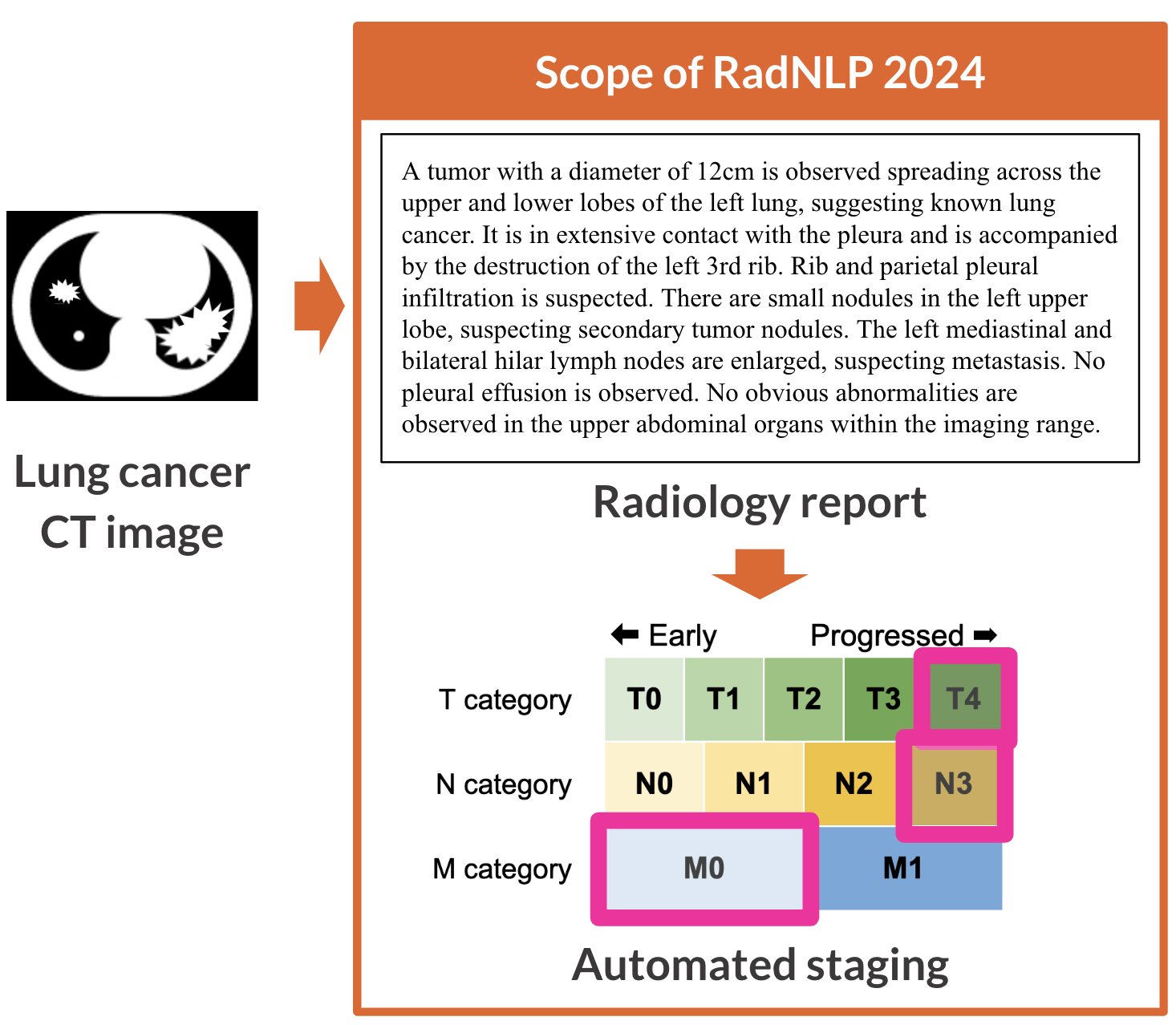
Sub task is a document segmentation to identify up to eight spans related to the following topics:
(i) Omittable – Spans that are clearly free of any positive findings or clearly unrelated to lung cancer staging. In this definition, “clearly” means the high clarity that does not require detailed knowledge of the lung cancer staging criteria.
(ii) Measure – Span describing mainly the existence and diameter of the primary lesion.
(iii) Extension – Span describing the range of the primary lesion’s extension outside the lung parenchyma.
(iv) Atelectasis – Span pointing out atelectasis or obstructive pneumonia.
(v) Satellite – Span pointing out intrapulmonary metastasis or lymphangiomatosis carcinomatosa.
(vi) Lymphadenopathy – Span pointing out enlarged regional lymph nodes.
(vii) Pleural – Span pointing out pleural/pericardial effusion/dissemination.
(viii) Distant – Span pointing out distant metastasis outside the lung parenchyma.
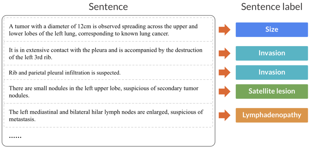
In NLP terminology, this sub task is a multi-label sentence binary classification.
The segmentation is at the sentence level, and it is possible for a topic span to be discontinuous or for the same sentence to belong to more than one topic.
Participants are requested to determine whether each sentence falls into categories (i) to (viii). If it does, mark it as “1,” and if it does not, mark it as “0.” Therefore, each sentence requires eight binary answers.
Note that every sentence will either fall into only category (i) Omittable, or into one or more of categories (ii) Measure to (viii) Distant.
In RadNLP 2024, we follow the staging criteria by the Japan Lung Cancer Society (JLCS). Note that the JLCS criteria closely aligns the global standard, namely the 8th edition of the TNM Classification of Malignant Tumours by the Union for International Cancer Control (UICC).
All radiology reports in RadNLP 2024 diagnose lung cancer at the initial evaluation. There are no reports of lung cancer post-treatment evaluation or different cancer.
Our datasets contain NO personal health information. The radiology reports are not derived from real medical institutions but are created with crowdsourcing by diagnosing de-identified images on Radiopaedia. Task participants requires no complex applications to use our datasets.
📊 Dataset Size & Format
This repository contains 6 files:
| Language | Split | Number of radiology reports | Number of sentences |
|---|---|---|---|
| English | Training | 108 | 918 |
| English | Validation | 54 | 415 |
| English | Test | 81 | 568 |
| English | Total | 243 | 1,901 |
| Japanese | Training | 108 | 1,020 |
| Japanese | Validation | 54 | 451 |
| Japanese | Test | 216 | 1,976 |
| Japanese | Total | 378 | 3,447 |
Each file contains 5 columns:
| Column | Type | Description |
|---|---|---|
id |
int | Unique identifier |
sentence_index |
int | Sentence index |
text |
string | Radiology Report |
omittable |
int | Gold standard for the "omittable" category |
measure |
int | Gold standard for the "measure" category |
extension |
int | Gold standard for the "extension" category |
atelectasis |
int | Gold standard for the "atelectasis" category |
satellite |
int | Gold standard for the "satellite" category |
lymphadenopathy |
int | Gold standard for the "lymphadenopathy" category |
pleural |
int | Gold standard for the "pleural" category |
distant |
int | Gold standard for the "distant" category |
📖 Citation
If you find our work useful, please consider citing:
@inproceedings{radnlp2024,
  title={NTCIR-18 RadNLP 2024 Overview: Dataset and Solutions for Automated Lung Cancer Staging},
  author={Nakamura, Yuta and Fujimoto, Koji and Kluckert, Jonas and Krauthammer, Michael and Kanzawa, Jun and Katayama, Akira and Kikuchi, Tomohiro and Kurokawa, Ryo and Gonoi, Wataru and Tashiro, Yuki and Hanaoka, Shouhei and Yada, Shuntaro and Aramaki, Eiji},
  year={2025},
  booktitle={NTCIR-18: Proceedings of the 18th NTCIR Conference on Evaluation ofInformation Access Technologies},
  doi={10.20736/0002002061}
}
📜 License
This project is licensed under the CC-BY-4.0 license.
- Downloads last month
- 18